Anatomy Diagram Rib Area : Rib Cage Stock Photos Stock Images And Vectors Stockfresh
Anatomy Diagram Rib Area : Rib Cage Stock Photos Stock Images And Vectors Stockfresh. The ribs are a set of twelve paired bones which form the protective 'cage' of the thorax. Human anatomy for muscle, reproductive, and skeleton. The rib cage is a bony structure found in the chest (thoracic cavity). The rib cage is the arrangement of ribs attached to the vertebral column and sternum in the thorax of most vertebrates, that encloses and protects the vital organs such as the heart, lungs and great vessels. We hope this picture human rib anatomy in detail can help you study and research.
ads/bitcoin1.txt
Rib cage anatomy the rib cage, shaped in a mild cone shape and more flexible than most bone sets, is made up of varying elements such as the thoracic vertebra, 12 equally paired ribs, costal cartilage, and held together anteriorly by the sternum. The primary responsibilities of the ribcage involve protecting the thoracic visceral organs, enclosing the thoracic visceral organs, and is included. Interactive tutorials about the ribs and sternum bones, with labeled images and diagrams featuring the beautiful illustrations of getbodysmart. Ribs sternum rib cage diagram function anatomy. We hope this picture human rib anatomy in detail can help you study and research.
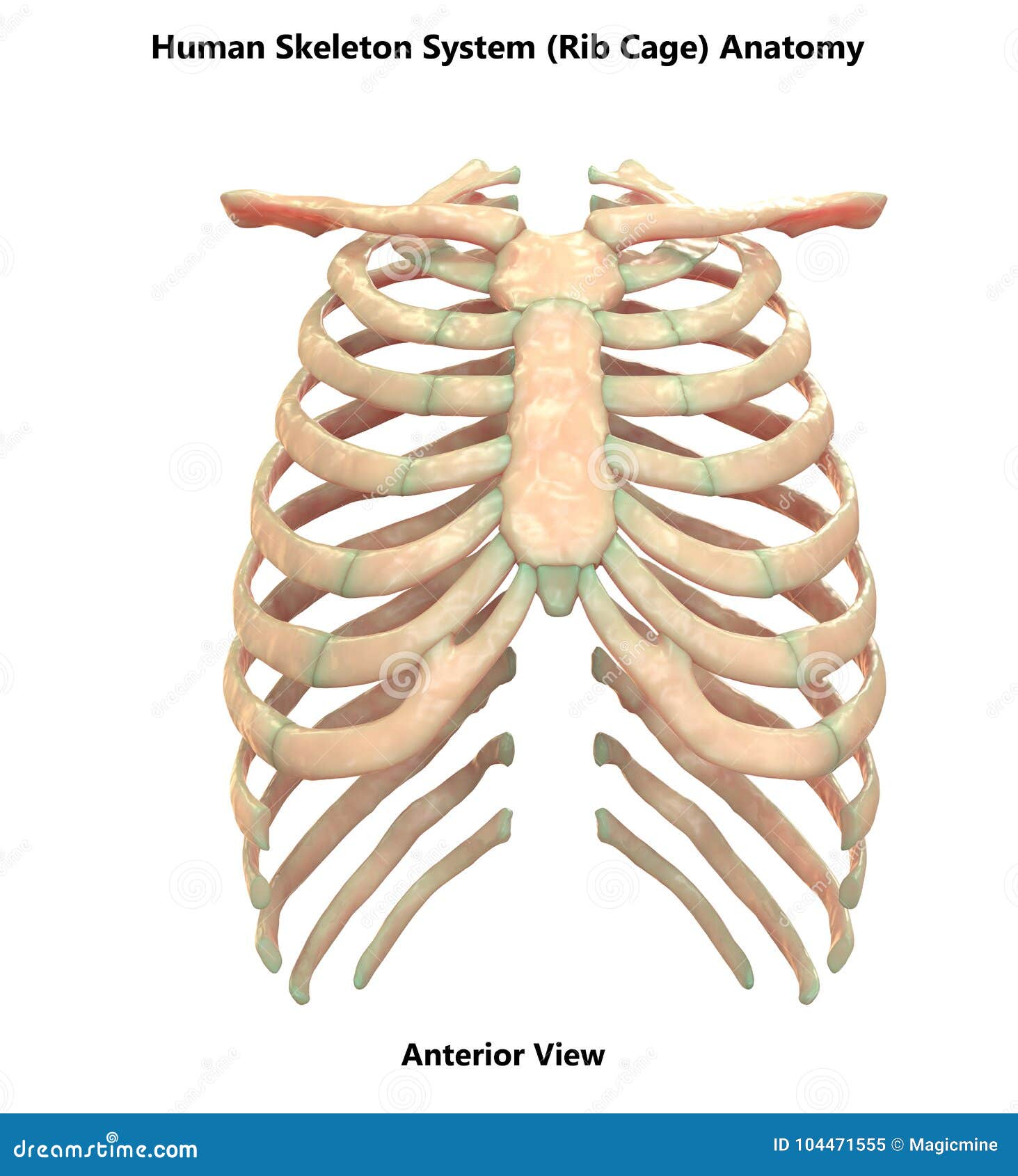
Human Skeleton System Rib Cage Anterior View Anatomy Stock Illustration Illustration Of Health Arthritis 104471555 from thumbs.dreamstime.com We hope this picture human rib anatomy in detail can help you study and research. Diagram of the chest most cases of covid 19 where the person has been vaccinated have been. These muscles help the body bend at the waist. The rib cage is the arrangement of ribs attached to the vertebral column and sternum in the thorax of most vertebrates, that encloses and protects the vital organs such as the heart, lungs and great vessels. The head only articulates with the body of the t1 vertebra and therefore only one articulatory surface is present. Related posts of rib cage diagram with organs anatomy of human stomach. The human rib cage is made up of 12 pairs of ribs, some of which attach to a bony. The muscles of the abdomen protect vital organs underneath and provide structure for the spine.
Find out more about the individual muscles within the our engaging videos, interactive quizzes it forms the bulk of the chest area and can be easily seen on the surface in some people.
ads/bitcoin2.txt
These muscles help the body bend at the waist. Ribs 11 and 12 do not have necks or tubercles and the anterior. Anatomy diagram rib area : Anatomy of ribs and its related area, find out more about anatomy of ribs and its related area. Rib 1 is also flattened horizontally. Rib cage, in vertebrate anatomy, basketlike skeletal structure that forms the chest, or thorax, and is made up of the ribs and their corresponding attachments to the sternum (breastbone) and the vertebral column.the rib cage surrounds the lungs and the heart, serving as an important means of bony protection for these vital organs.in total, the rib cage consists of the 12 thoracic vertebrae and. We hope this picture human rib anatomy in detail can help you study and research. The superior surface is unique in that it is marked by two grooves that allow. The most common cause of sharp or dull pains in your rib cage is a pulled muscle. The primary responsibilities of the ribcage involve protecting the thoracic visceral organs, enclosing the thoracic visceral organs, and is included. The human rib cage is made up of 12 pairs of ribs, some of which attach to a bony. Rib 2 is thinner and longer than rib 1, and has two articular facets on the head as normal. By printing out this quiz and taking it with pen and paper creates for a.
The cartilage that forms at the end of each rib (costal cartilage) attaches either. The rib cage is collectively made up of long, curved individual. Anatomynote.com found human rib anatomy in detail from plenty of anatomical pictures on the internet. Anatomy of ribs and its related area, find out more about anatomy of ribs and its related area. Rib 1 is also flattened horizontally.
Rib Cage Anatomy Function Britannica from cdn.britannica.com Anatomy diagram rib area / anatomy of the female abdomen and pelvis, cut away view. In most tetrapods, ribs surround the chest, enabling the lungs to expand and thus facilitate breathing by expanding the chest cavity. The ribs partially enclose and protect the chest cavity, where many vital organs (including the heart and the lungs) are located. The upper chest has two main functions: Ribs sternum rib cage diagram function anatomy. Learn vocabulary, terms and more with flashcards, games and other study tools. It is made up of 12 pairs of ribs. We think this is the most useful anatomy picture that you need.
For more anatomy content please follow us and visit our website:
ads/bitcoin2.txt
The rib cage is the arrangement of ribs attached to the vertebral column and sternum in the thorax of most vertebrates, that encloses and protects the vital organs such as the heart, lungs and great vessels. The rib cage is an important part of the human anatomy. The major muscles of the abdomen include the rectus. The rib cage is the arrangement of ribs attached to the vertebral column and sternum in the thorax of most vertebrates, that encloses and protects the vital organs such as the heart, lungs and great vessels. The upper chest has two main functions: Anatomy diagram rib area / pin on anatomy : The ribs partially enclose and protect the chest cavity, where many vital organs (including the heart and the lungs) are located. Human anatomy for muscle, reproductive, and skeleton. Anatomy diagram rib area / pin on anatomy : The first rib is the widest, shortest and has the sharpest curve of all the ribs. The primary responsibilities of the ribcage involve protecting the thoracic visceral organs, enclosing the thoracic visceral organs, and is included. The cartilage that forms at the end of each rib (costal cartilage) attaches either. Find out more about the individual muscles within the our engaging videos, interactive quizzes it forms the bulk of the chest area and can be easily seen on the surface in some people.
Anatomical planes of the body. The major muscles of the abdomen include the rectus. Ribs sternum rib cage diagram function anatomy. The ribs partially enclose and protect the chest cavity, where many vital organs (including the heart and the lungs) are located. Check out the premium anatomy course to see the full version of this video and all other anatomy videos.
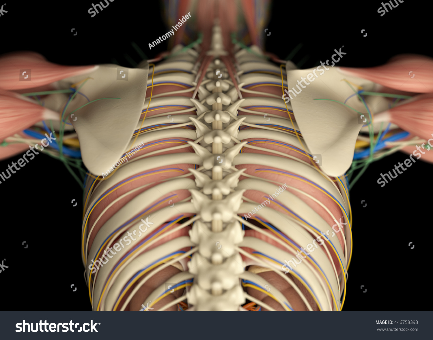
Human Anatomy Back Rib Cage Torso Stock Illustration 446758393 from image.shutterstock.com The heads of ribs 1, 10, 11, and 12 have a single facet for articulation with the bodies of the thoracic vertebrae. As viewed from the side, the thoracic spine's vertebrae form a kyphotic curve that runs from t1 to t12, in which the spine curves outward towards the back of the body to allow. The rib cage is a bony structure found in the chest (thoracic cavity). For more anatomy content please follow us and visit our website: It has a roughened area on its upper surface, from which the serratus anterior muscle originates. As in the typical ribs, the tubercle has a facet for articulation with the transverse process of vertebrae. Related posts of rib cage diagram with organs anatomy of human stomach. Anatomy diagram rib area :
The thoracic cage is a component of the thoracic wall and encloses the majority of the structures of the respiratory system.
ads/bitcoin2.txt
Rib cage diagram | healthiack / vector art, clipart and stock vectors. The rib cage is an important part of the human anatomy. Learn vocabulary, terms and more with flashcards, games and other study tools. These muscles help the body bend at the waist. Anatomy diagram rib area / pin on anatomy : The most common cause of sharp or dull pains in your rib cage is a pulled muscle. The anatomy of the human ribs is made up of 24 ribs which are parted in 12 pairs (each on the left and right side of the chest wall), with the sternum, metasternum (the xiphoid process), and the costal cartilages all situated at the anterior of the chest wall, followed by the thoracic vertebrae on the posterior of the chest wall. This bony framework plays an essential role in protecting the organs that lie in the thoracic region. By printing out this quiz and taking it with pen and paper creates for a. Numbered ribs, sternum, cartilage parts and clavicular articulation.ribs eight to ten are the false ribs and are connected to the sternum indirectly via the cartilage of the rib above learn everything about the ribs with our articles, video tutorials, quizzes, and labeled diagrams there are eleven pairs of external intercostal muscles and these are. Anatomy diagram rib area : It has a roughened area on its upper surface, from which the serratus anterior muscle originates. The first rib is the widest, shortest and has the sharpest curve of all the ribs.
ads/bitcoin3.txt
ads/bitcoin4.txt
ads/bitcoin5.txt
0 Response to "Anatomy Diagram Rib Area : Rib Cage Stock Photos Stock Images And Vectors Stockfresh"

0 Response to "Anatomy Diagram Rib Area : Rib Cage Stock Photos Stock Images And Vectors Stockfresh"
Post a Comment